The Gold Standard for Bacterial Quantification
Colony forming unit (CFU) testing represents the gold standard method for measuring bacterial populations in samples ranging from cleaning product efficacy studies to food safety testing. Understanding CFU testing reveals how scientists quantify bacterial reductions, validate cleaning product claims, and ensure safety in various applications. This fundamental microbiological technique bridges laboratory precision with real-world bacterial control, providing the numerical evidence supporting claims that products reduce bacteria by specific percentages.
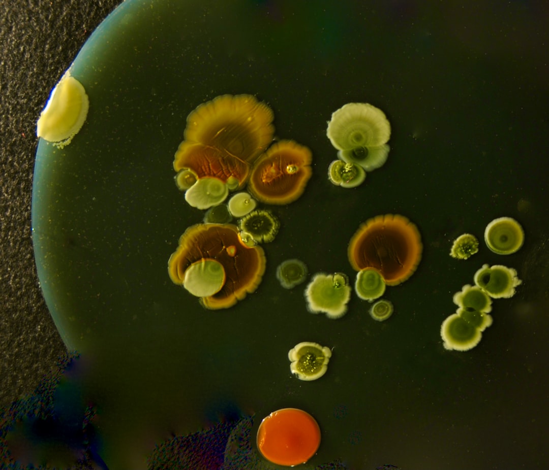
The principle behind CFU testing proves elegantly simple: dilute a bacterial sample sufficiently that individual bacteria separate, spread the diluted sample on nutrient agar, incubate to allow bacterial growth, and count the resulting colonies. Each visible colony theoretically represents one viable bacterium from the original sample—one colony-forming unit. Multiply the colony count by the dilution factor, and you obtain an estimate of viable bacterial concentration in the original sample.
The CFU Testing Process
CFU testing begins with sample collection. For surface testing, sterile swabs or sponges collect bacteria from defined surface areas—typically 25 or 100 square centimetres. The swab or sponge is then placed in sterile buffer solution, and vigorous mixing releases bacteria into the liquid. For liquid samples like cleaning products or food rinses, samples are used directly or after appropriate dilution.
Serial dilution creates the manageable bacterial concentrations necessary for counting. Undiluted samples typically contain far too many bacteria to count individual colonies—they'd create confluent growth covering entire plates. Serial dilutions involve transferring small volumes (often 1 mL or 0.1 mL) into larger volumes (9 mL or 9.9 mL) of sterile buffer, creating 10-fold or 100-fold dilutions. Repeating this process multiple times generates dilution series: 10-1, 10-2, 10-3, and so forth.
Plating involves spreading specific volumes from various dilutions onto nutrient agar plates. The spread plate technique uses sterile spreaders to distribute samples evenly across agar surfaces. The pour plate method mixes samples with molten agar before pouring into plates, embedding bacteria throughout the agar. Both techniques work; different laboratories prefer one or the other based on tradition and specific applications.
Incubation allows bacterial growth under controlled conditions. For most environmental bacteria, incubation at 30-37°C for 24-48 hours proves sufficient. Specific organisms might require different conditions: some prefer cooler temperatures, others need extended incubation, and some require specialised nutrient media. Proper incubation transforms invisible individual bacteria into visible colonies that can be counted.
Counting and Calculation
After incubation, trained technicians count colonies on plates. For accurate counting, plates should contain 30-300 colonies—fewer provides poor statistical precision, whilst more creates overlapping colonies that prove difficult to distinguish. This requirement explains why multiple dilutions are plated; at least one dilution should yield countable numbers.
The calculation converts colony counts into CFU per millilitre (for liquids) or CFU per square centimetre (for surfaces). The formula accounts for dilution factor and plated volume:
CFU/mL = (Number of colonies × Dilution factor) / Volume plated
For example: if a 10-4 dilution plated at 0.1 mL yields 85 colonies, the calculation proceeds: 85 × 10,000 (dilution factor) / 0.1 = 8.5 × 10⁶ CFU/mL. This means the original sample contained 8.5 million viable bacteria per millilitre.
Measuring Cleaning Product Effectiveness
CFU testing validates cleaning product antimicrobial claims by comparing bacterial counts before and after product application. Standard protocols involve contaminating test surfaces with known bacterial concentrations, applying cleaning products according to manufacturer instructions, sampling surfaces after specified contact times, and quantifying surviving bacteria via CFU counting.
The percentage reduction calculation compares treated and untreated surfaces:
% Reduction = [(CFUcontrol - CFUtreated) / CFUcontrol] × 100
Products claiming "99.9% bacterial reduction" must demonstrate that treated surfaces contain 1,000-fold fewer bacteria than untreated controls. For instance, if control surfaces average 106 CFU/cm², treated surfaces must average ≤103 CFU/cm². This dramatic reduction represents what regulatory agencies and consumers expect from antimicrobial products.
Testing multiple bacterial species ensures broad-spectrum effectiveness. Standard test organisms include Escherichia coli (Gram-negative), Staphylococcus aureus (Gram-positive), and sometimes Pseudomonas aeruginosa or Salmonella species. Products should demonstrate specified reductions against all test organisms to claim broad-spectrum activity.
Probiotic Product Testing Considerations
Testing probiotic cleaning products requires modified approaches because these products contain live bacteria that will appear in CFU counts. Standard antimicrobial testing assumes product bacteria either don't grow on test media or can be distinguished from challenge organisms. Probiotic bacteria complicate this assumption.
Selective media partially address this challenge. If testing probiotic products against E. coli, using MacConkey agar (which selectively grows Gram-negative bacteria whilst inhibiting Gram-positives like Bacillus) allows counting E. coli whilst excluding probiotic bacteria. Similarly, mannitol salt agar selectively grows Staphylococcus species, allowing S. aureus quantification despite probiotic presence.
However, complete selectivity proves challenging. Some modified protocols use paired sampling: one sample plated on non-selective medium (counting all bacteria) and another on selective medium (counting only challenge organisms). The difference estimates probiotic bacterial populations, whilst selective counts reveal challenge organism reductions.
Molecular methods like quantitative PCR can distinguish bacterial species based on genetic markers, allowing probiotic and challenge organism quantification in mixed populations. Whilst more complex than traditional CFU testing, these methods provide accurate data for probiotic product validation.
Time-Kill Studies
Time-kill studies extend basic CFU testing by measuring bacterial reductions at multiple time points, revealing how quickly products achieve antimicrobial effects. Samples are collected at 0, 5, 10, 30, 60 minutes, and longer intervals post-treatment, with CFU quantification at each time point.
Chemical disinfectants typically show rapid bacterial reductions—achieving maximum effects within minutes. Time-kill curves for disinfectants drop steeply initially then plateau as surviving resistant bacteria persist. Probiotic products show different kinetics: modest initial reductions followed by progressive decreases over hours as beneficial bacteria establish populations and competitive effects manifest.
These temporal patterns explain why testing protocols matter. Studies evaluating only immediate effects (within minutes or hours) favour chemical disinfectants. Including extended time points (24-72 hours) reveals probiotic advantages in sustained bacterial suppression.
Surface Carrier Testing
Surface carrier tests assess product performance on realistic surfaces rather than in suspension. Standard methods use stainless steel, ceramic, or plastic carriers contaminated with bacteria, dried to mimic natural contamination, treated with products, and sampled for CFU quantification.
Surface testing proves more challenging than suspension testing because bacteria dry onto surfaces, creating adhesion that resists removal. Sampling techniques must recover attached bacteria without introducing excessive variability. Standardised protocols specify swabbing or rinsing methods that provide reproducible bacterial recovery from surfaces.
Results from surface carrier tests often differ from suspension tests, typically showing lower bacterial reductions because surface attachment protects bacteria. Products might achieve 99.99% reduction in suspension but only 99% on surfaces, highlighting the importance of testing under realistic conditions.
Biofilm CFU Quantification
Quantifying bacteria in biofilms requires disrupting biofilm matrices to release embedded bacteria whilst maintaining their viability for CFU counting. Sonication applies ultrasonic vibrations that physically disrupt biofilms without killing bacteria. Vortexing with glass beads mechanically breaks up biofilms. Enzymatic treatment degrades biofilm matrices, releasing bacteria.
After biofilm disruption, standard serial dilution and plating proceeds. Comparing CFU counts from biofilm samples before and after treatment reveals product effectiveness against biofilm bacteria. These tests prove particularly relevant for probiotic products, which claim biofilm degradation through enzyme production—a claim CFU testing can validate.
Statistical Considerations
CFU counts show inherent variability from multiple sources: sampling variability, plating variability, and biological variability in bacterial populations. Proper experimental design addresses this through replication—testing multiple samples at each condition—and statistical analysis comparing means whilst accounting for variability.
Results are typically reported as mean CFU counts with standard deviations or standard errors. Statistical tests (t-tests, ANOVA) determine whether observed differences between treatments prove statistically significant rather than random variation. Regulatory acceptance of antimicrobial claims often requires demonstrating statistically significant reductions with specified confidence levels.
Limitations of CFU Testing
Despite being the gold standard, CFU testing has limitations. It quantifies only culturable bacteria—those able to grow on provided media under specified conditions. Viable but non-culturable (VBNC) bacteria, damaged bacteria that might recover, or fastidious bacteria requiring specialised growth conditions won't appear in CFU counts.
CFU testing provides no information about bacterial identity beyond what selective media reveal. Without additional characterisation, technicians cannot distinguish between pathogenic and beneficial bacteria if both grow on test media. Molecular methods or biochemical testing must supplement CFU counting for species identification.
The technique requires 24-48 hours for results, limiting real-time decision making. Rapid methods exist (ATP bioluminescence, flow cytometry) but don't provide the same detailed quantitative data as CFU testing.
Quality Control and Method Validation
Reliable CFU testing requires rigorous quality control. Laboratories must validate methods using reference strains with known characteristics, run positive and negative controls with each experiment, and participate in proficiency testing programmes comparing results across laboratories.
Media quality control ensures nutrient agar supports expected bacterial growth. Known bacterial concentrations plated on each media batch verify fertility. Sterility testing confirms media aren't contaminated. Equipment calibration—incubators maintaining accurate temperatures, pipettes delivering precise volumes—proves essential for reproducible results.
Trained personnel represent perhaps the most critical quality control element. Colony counting requires skill recognising colonies versus background contamination or media artifacts. Experienced technicians produce more reliable data than untrained staff, explaining why reputable testing laboratories invest heavily in personnel training.
Interpreting CFU Data
Understanding CFU test results requires context. A 99% bacterial reduction sounds impressive but leaves 1% surviving—potentially millions of bacteria on a heavily contaminated surface. Whether this proves acceptable depends on application: for surgical instrument sterilisation, 99% proves insufficient; for routine household cleaning, it may provide adequate safety margins.
Comparing products requires examining complete data including: bacterial species tested, surface types, contact times, environmental conditions, and statistical analyses. Claims like "kills 99.9% of germs" mean little without specifications. Which germs? Under what conditions? Within what time frame? Complete CFU testing data answers these questions, enabling informed product comparison.
The Future: Complementary Methods
Whilst CFU testing will remain fundamental, complementary methods provide additional insights. Quantitative PCR measures total bacterial DNA, including non-culturable cells. Flow cytometry rapidly sorts living from dead bacteria. ATP bioluminescence quantifies metabolic activity. Combining multiple methods creates comprehensive bacterial quantification accounting for CFU testing's limitations.
For probiotic product testing, multi-method approaches prove particularly valuable. CFU testing quantifies viable culturable bacteria, molecular methods identify and quantify specific species, and microscopy visualises surface colonisation patterns. Together, these techniques thoroughly validate probiotic cleaning effectiveness, providing the robust evidence supporting this innovative approach to bacterial management.